 EngEng
On the basis of the surgical department of the University Clinic at Tinista, 8 the latest technologies for the treatment of patients with malignant skin tumors have been introduced.
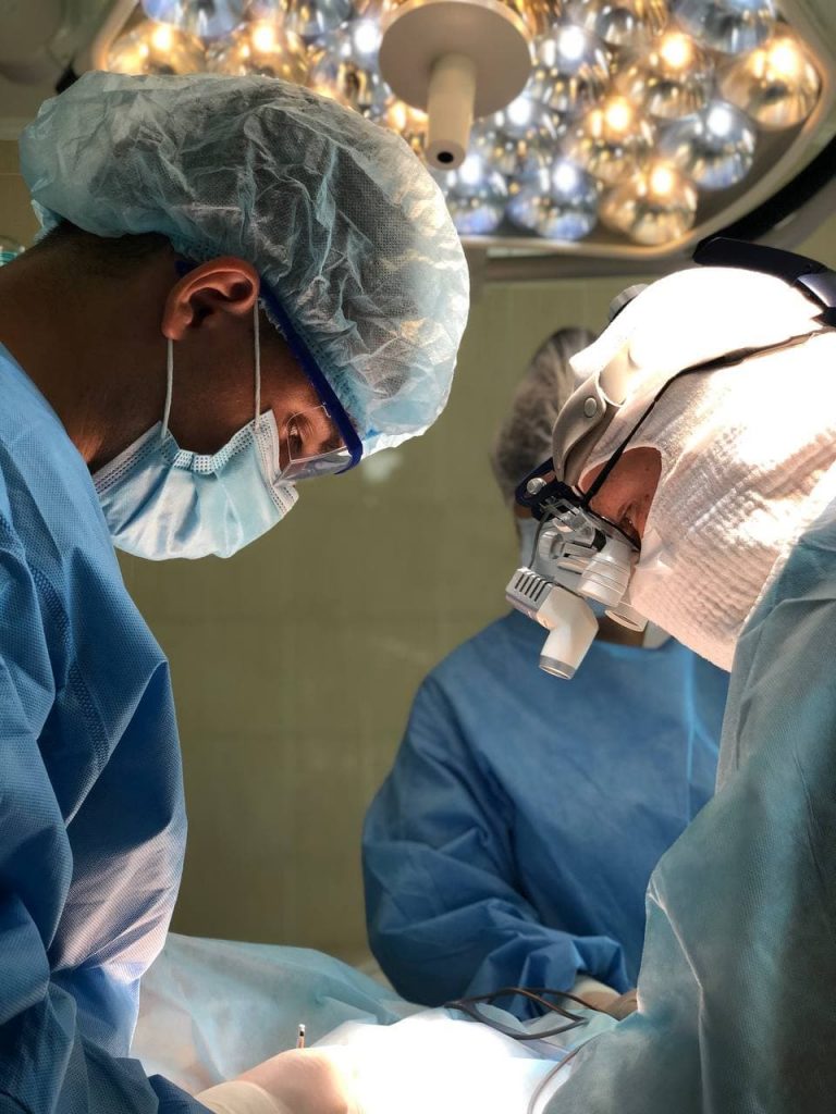
Modern equipment is used in accordance with international treatment protocols at all stages of treatment. Mohsmicrographic surgery is used for patients with basal cell and squamous cell carcinomas of the skin. Today, Mohs micrographic surgery is included in all European and American protocols for the treatment of non-melanoma skin cancers.
The main difference between Mohs surgery and classical surgical excision is that the tumor is removed in stages, with histological control of the boundaries after each stage and determining the spread of the tumor in the tissue with reference to 3D spatial orientation. Thanks to a special technique, Mohs surgery provides complete removal of the tumor with minimal involvement of the surrounding healthy tissues, which allows to minimize the surgical defect and get the best aesthetic result.
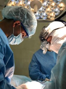
Doctors of the surgical department perform a full range of plastic and reconstructive operations to close the defects after excision of malignant skin tumors.
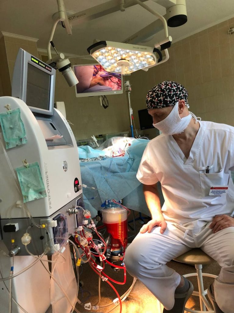
Isolated hyperthermic chemoperfusion of the extremities (ILP) is used for patients with locally advanced melanoma of the skin of the extremities when surgery cannot be performed.
The ILP method was developed by Krich and Kremenets in 1956 to achieve high concentrations of chemotherapeutic drug in the extremity affected by inoperable tumors, especially soft tissue sarcoma and melanoma, and to minimize toxicity associated with systemic chemotherapy. For this purpose, the blood circulation of the affected limb is isolated from the systemic circulation and connected to the extracorporeal system. After reaching high temperatures, the patient is administered chemotherapeutic drugs through the perfusion circuit, mainly melphalan and tumor necrosis factor (TNF). Studies show satisfactory data on relapse-free and overall survival using this method. ILP also has a low level of regional and systemic toxicity compared to systemic chemotherapy.
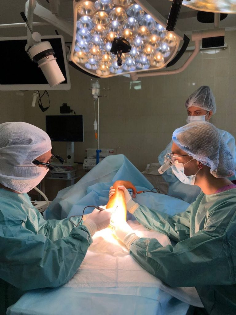
On the basis of the surgical department, a biopsy of sentinel lymph nodes is performed under fluoroscopic control. The essence of sentinel lymph node biopsy is that there is a primary, or sentinel, lymph node (or nodes) through which tumor cells from the primary tumor at a particular location must first pass to enter a specific regional lymph node pool. The indicator substance (indocyanine green), injected into the dermis at the site of the primary tumor, provides a road map leading to the sentinel lymph nodes. Careful examination of the signaling lymph nodes indicates the condition of the entire lymph node basin, which has been confirmed in many international studies. Thus, sentinel lymph node biopsy with selective lymphadenectomy has been adopted as an alternative to routine lymphadenectomy or follow-up of patients with clinically negative regional lymph nodes who are at risk for nodular metastases.
To prevent damage to nerve structures, working in anatomically complex areas, the surgical department of the University Clinic uses intraoperative neuromonitoring, which allows you to accurately identify and preserve nerve structures. At germination by a tumor of the main vessels vascular surgeons who carry out prosthetics of any complexity are involved in work. A multidisciplinary team of specialists guarantees the patient highly specialized care.

 Ukr
Ukr