 EngEng
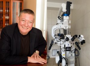
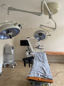
Neurosurgeon is one of the most difficult medical disciplines. One wrong move by a surgeon can result in a disability for the patient. That is why safety measures during operations on the brain and spinal cord are absolutized. The most complex neurosurgical operations are performed in the university clinic of Odessa National Medical University. Recently, a unique neurosurgical operating microscope was purchased for the clinic, which has no analogues in Ukraine. About modern neurosurgical technologies and equipment that made them available in our country, – told us in an interview the head of the Department of Neurology and Neurosurgery of Odessa National Medical University, Professor Anatoliy Son.
Tell us about the microscope. What opportunities does it give to doctors and patients?
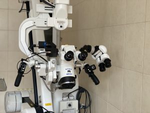
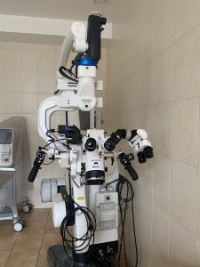
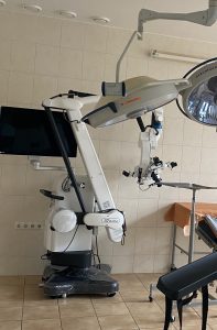
We can say that there are two such models in the world. One – from a German manufacturer, the other – the Japanese Mitaka. They are equal in many respects, both have modules that allow you to contrast the tumor or perform angiography – to see the vessels during surgery. But Mitaka, which now stands at the University Clinic, has an important advantage – better depth of field.
We work in a deep narrow wound, at the bottom of which there is an aneurysm or a tumor. For better work it is necessary to see the whole wound equally well. This is the depth of field. Most microscopes allow us to see well only what we do directly – remove the tumor or clip the aneurysm. We see the rest of the wound badly and indistinctly. Our microscope solves this problem. It’s like comparing what’s better: stereoscopic vision or planar vision. The operating microscope makes work much easier, makes movements more accurate.
What magnification does it give?
This is the only operating microscope in the world that gives a magnification of more than 70. They even write that the maximum is 72-74. This is an unprecedented increase. The rest of the microscopes give up to 50.
Is there a need for such an increase during operations?
For example, we apply microanastomoses on vessels, sew vessels with a diameter of less than 1 mm. To effectively suture a vessel and, most importantly, to make it work afterwards, we need to see the vessel wall well. This requires a large increase. Yes, not necessarily 70 times. We often work at a lower magnification, but we get much better and clearer images than with a conventional operating microscope. This is a very important advantage.
The mobility of the microscope is also important for neurosurgeons. For example, an ophthalmologist changes the lens. He pointed the microscope – and the device is in this position throughout the operation. We need to constantly change the angle of view during operations. The suspension system allows you to change the angle of the view at any time, while the microscope does not overlook the area we need.
Any microscope accelerates operations and increases their accuracy. What to say about a microscope with such functionality. I have been working as a neurosurgeon since 1978, for more than 40 years. Of these, since 1985 I have operated only with a microscope – most of my life. When I came to Odessa, 25 years ago, neurosurgeons did not operate with a microscope here. My colleagues told me: “Let’s operate without a microscope, it will be faster.” Once they wre convinced that the microscope is faster and better, no operation was performed without it any more.
Does it allow you to operate on more difficult patients?
All our patients are difficult. Neurosurgery has long been exclusively microsurgery. Good microscopes allow you to see better, make more precise movements, spend less time. You see, the speed of the operation is not a goal in itself. The patient is under anesthesia. Undoubtedly, the less time he is under it – the better it is for him.
Is the Mitaka microscope expensive to maintain?
Let’s say we have a protective sterile cover, which we must put on the microscope during the operation. For operating microscopes from other manufacturers, this case costs approximately 25 euros and is disposable. We can use our Ukrainian covers for a conditional UAH 100. Why? Because in our microscope, protective glass is provided on the lens, and in other manufacturers – in each case has its own glass. I can say that it significantly reduces the cost of surgery and simplifies our lives.
All the 25 years in Odessa you work in a university clinic. How has it changed over the years?
Yes, all these years my workbook is in one place – at the university. But we have different clinical bases. In the first years I worked in a military hospital, then in a city hospital. At that time, the university clinic was not sufficiently equipped. Now all we have is ultra-modern.
What else but the microscope?
Each neurosurgeon has two instruments in his hands during the operation. This is an aspirator and tweezers for coagulation of blood vessels. A conventional aspirator removes fluid from the wound. For six months now, we have been using ultrasound, which, in addition, destroys the tumor itself without injuring nerves and blood vessels. This is top German equipment, absolutely invaluable in terms of the patient’s safety.
You see, the brain is a special organ. If we dissect it, it is a dysfunction. We need to dissect the brain. Firstly, you just can’t do it without a microscope. Secondly, without electrical coagulation. How does it work? We apply current to the tweezers – there is coagulation of blood vessels. The tweezers seem to burn and stick to the fabric. If we remove the tweezers from the fabric, it tears it. Now we have an electrosurgical system that allows you to operate without burning tweezers. Accordingly, the injury is reduced to zero. And all this gives the main thing – a high-quality and successful operation.
Operations on the brain differ from any other ones in that the team during the operation must constantly monitor its functions…
During operations, we monitor the functions of the brain and nerves with a special neuromonitoring system, which helps to constantly monitor their functions, and therefore, it is safe for the functions of the brain and nerves to remove the tumor. Our operating table is X-ray transparent. When we operate on the spine, X-ray control is performed all the time. Do you understand what an X-ray transparent table means to us? Or, say, during brain surgery, we rigidly attach the head to the table. This must be required. Sometimes we wake patients to see how the brain works. At the same time, not the slightest movement can be allowed, so the head is fixed. This is called a headholder. It is also radiopaque. It would seem, why is this necessary? Our plans include intraoperative computed tomography, which can only be done with a transparent headholder. What it is? During the operation, we must monitor the completeness of tumor removal. It looks something like this: somewhere in the corner of the operating room there is a portable computer tomograph. We press a button – it moves to the table, “comes” to the patient’s head and performs the tomography. Therefore, the headholder must be transparent to X-rays. Usually we do this after the operation – we take the patient to a large tomograph. Imagine how important it is to be able to control your actions during the surgery.
Are students trained on this equipment or is it used for clinical purposes only?
Of course, not all students have the opportunity to participate in operations. It is available to members of the scientific student group of our department. But all students must see how modern operations take place. Almost every one we broadcast for them online simply from an operating microscope or endoscope. Now in the conditions of distance learning, students are at home and can connect to the operation at any time. They need to see complex cases and understand how the neurosurgeon handles them.
Patient safety during operations on the nervous system is paramount. It cannot be guaranteed without equipment. Undoubtedly, qualified personnel is needed. But only people are not enough here. For neurosurgeons, the most important thing is brain function. Behind each of our movements during the operation there is a function of the brain. Poor quality work of neurosurgeons is very visible. The consequences of even the most difficult heart surgery are not visible visually. Everyone sees the mistakes of neurosurgeons: a person either does not see, or the hand is paralyzed, and so on. High qualification of the doctor in a complex with the top equipment can guarantee that such things will not happen.
 Ukr
Ukr