 EngEng
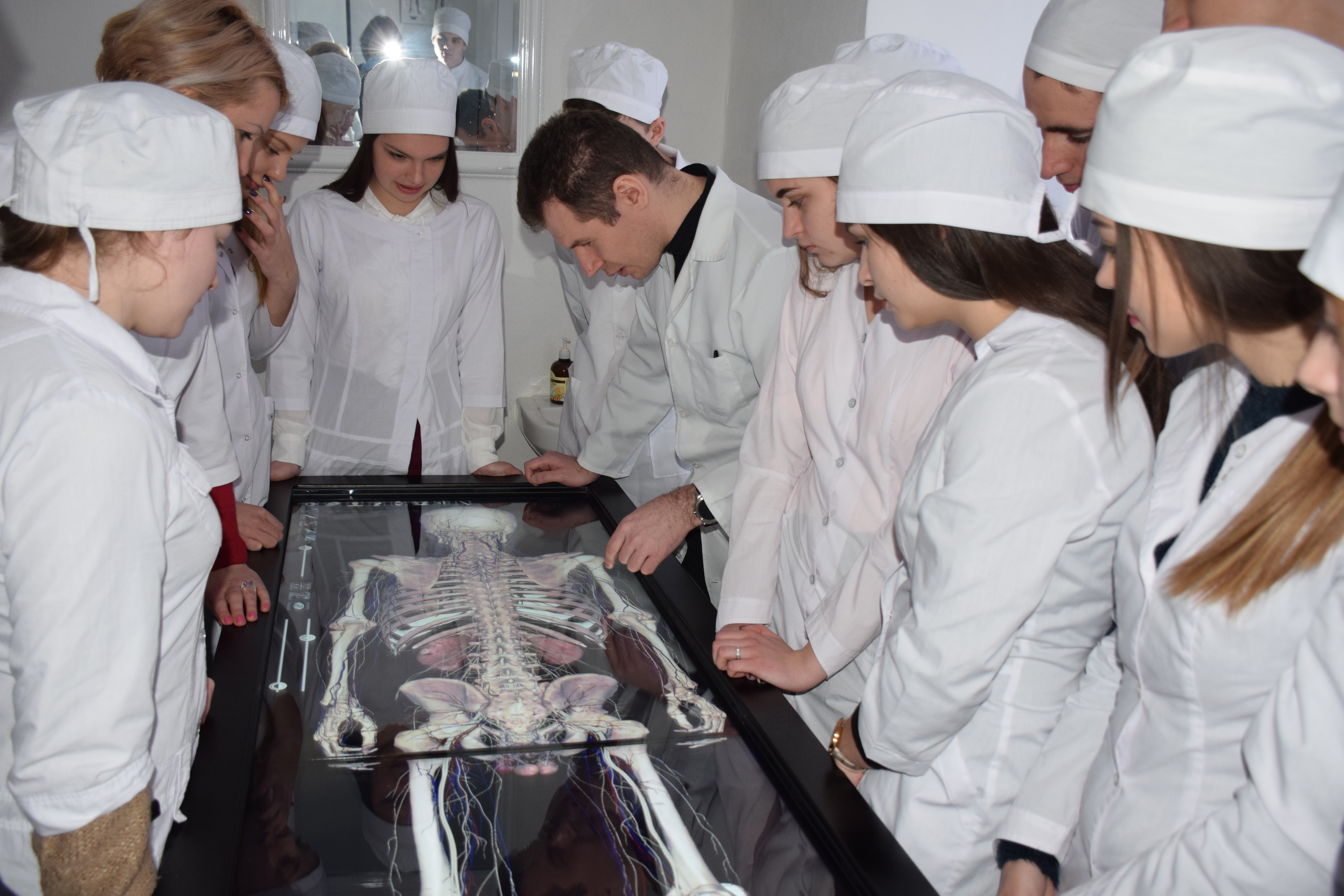
In April this year, the professors of the Department of Anatomy held scheduled lessons on current topics at the Department of Simulation Medicine. Students of medical faculties got acquainted with the work of the interactive computer table ANATOMAGE TABLE and possibilities of its use for studying human anatomy.
Students looked at the location of organs, vessels and nerves in 3D projections, and they figured out their topography. They also used the option of layer-wipe removal of tissues with a subsequent review of deep body structures. Using anatomical table during practical classes allowed to study the external and internal structure of the bodies macroscopically. All together it gave the effect of a better perception of the integrity of the organism and made the process of studying more interesting. It was possible to work out the method of virtual preparation.
In our view, the anatomical virtual table provides a more complete and effective study of the human anatomy. Working with virtual biological material allows us to evaluate the topography of the organ (skeletotopy, syntopy, and holotopy), its blood supply and innervation, and to understand the structure of the organism as a whole. The use of this teaching technique encourages not only students but also teachers, since high-tech equipment greatly improves and broadens knowledge of the discipline.
 Ukr
Ukr