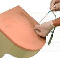
This ultrasound compatible model allows for procedural accuracy when performing the paracentesis – draining fluid from the peritoneal cavity in the abdomen. The anatomically correct model allows the procedure to be performed with or without ultrasound and on either of the recommended areas: the midline below the umbilicus or medial, 4 to 5 cm above the anterior superior iliac spine
FeaturesandBenefits:
- Allows for procedural accuracy
- Ultrasound compatible with replaceable tissue
- Anatomy includes superficial epigastric vessels, partial liver and partial spleen, rectus abdominal muscles, and mesentery intestines
- Anatomical landmarks include the pubis symphysis, iliac crest, and umbilicus
- Allows up to one liter of intraperitoneal fluid removal
- Replaceable tissue is durable and allows for repeated use
- Palpable anatomy and realistic needle response
 Eng
Eng Ukr
Ukr